Distal Ejecta Bed, SD02MI01
Location: Wickwar, near Bristol, England
|
Description
Spherules deposited in a cross-bedded limestone which are
believed to be the result of a meteorite impact. Initially
these spherules would have been formed of silica but have
since been replaced with glauconitic minerals.
|
Photo of hand specimen
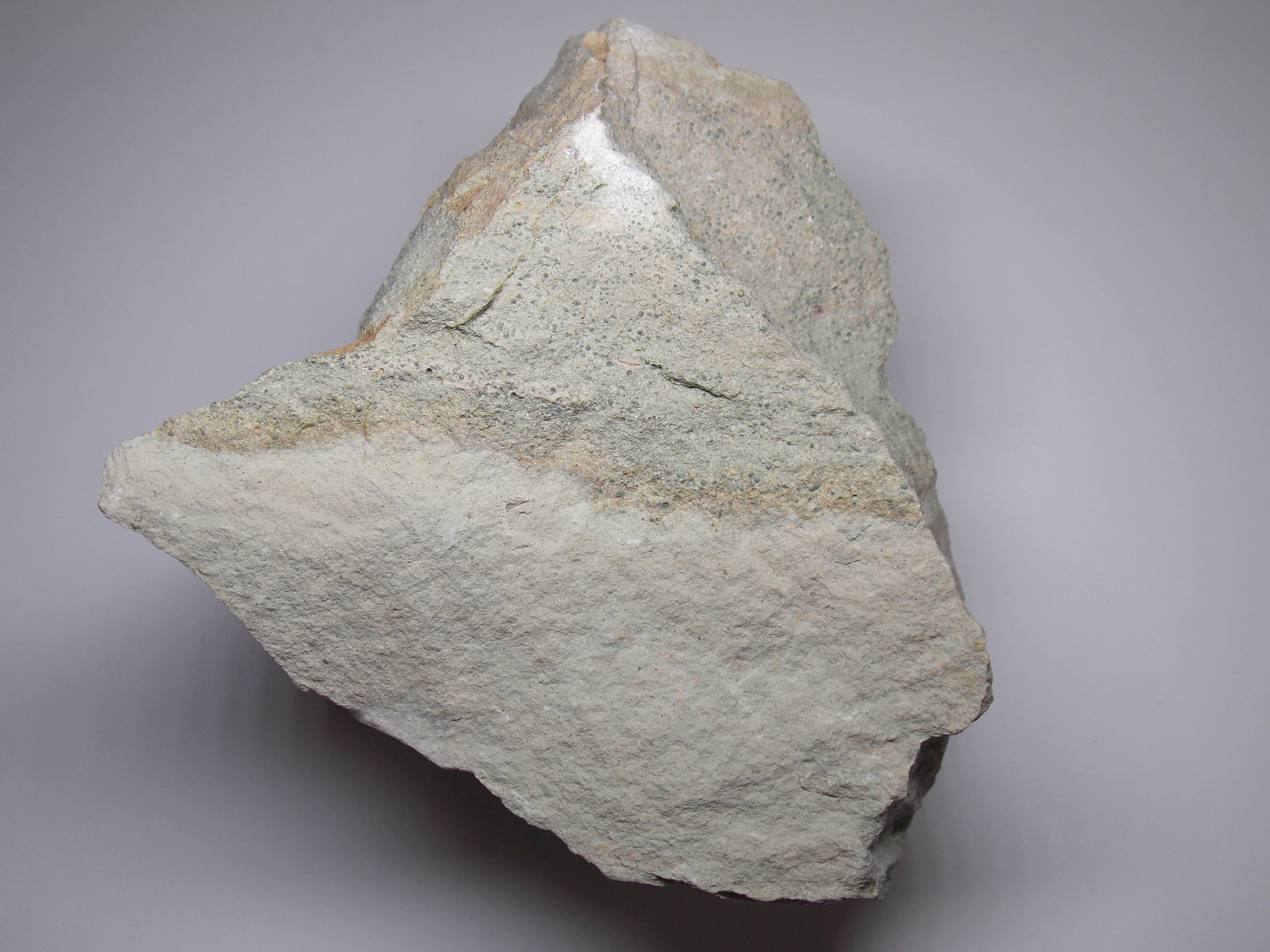
Specimen size: 110 x 100 x 50mm
Spherules can be seen in the upper portion of the
specimen. The spherules have been reworked into their
current position in a cross-bedded limestone and would
have originally been deposited in a thin layer over a
wider area. This later reworking has hampered attempts to
locate the original impact and the spherules are only
found in a geographically restricted area, largely
destroyed by quarrying.
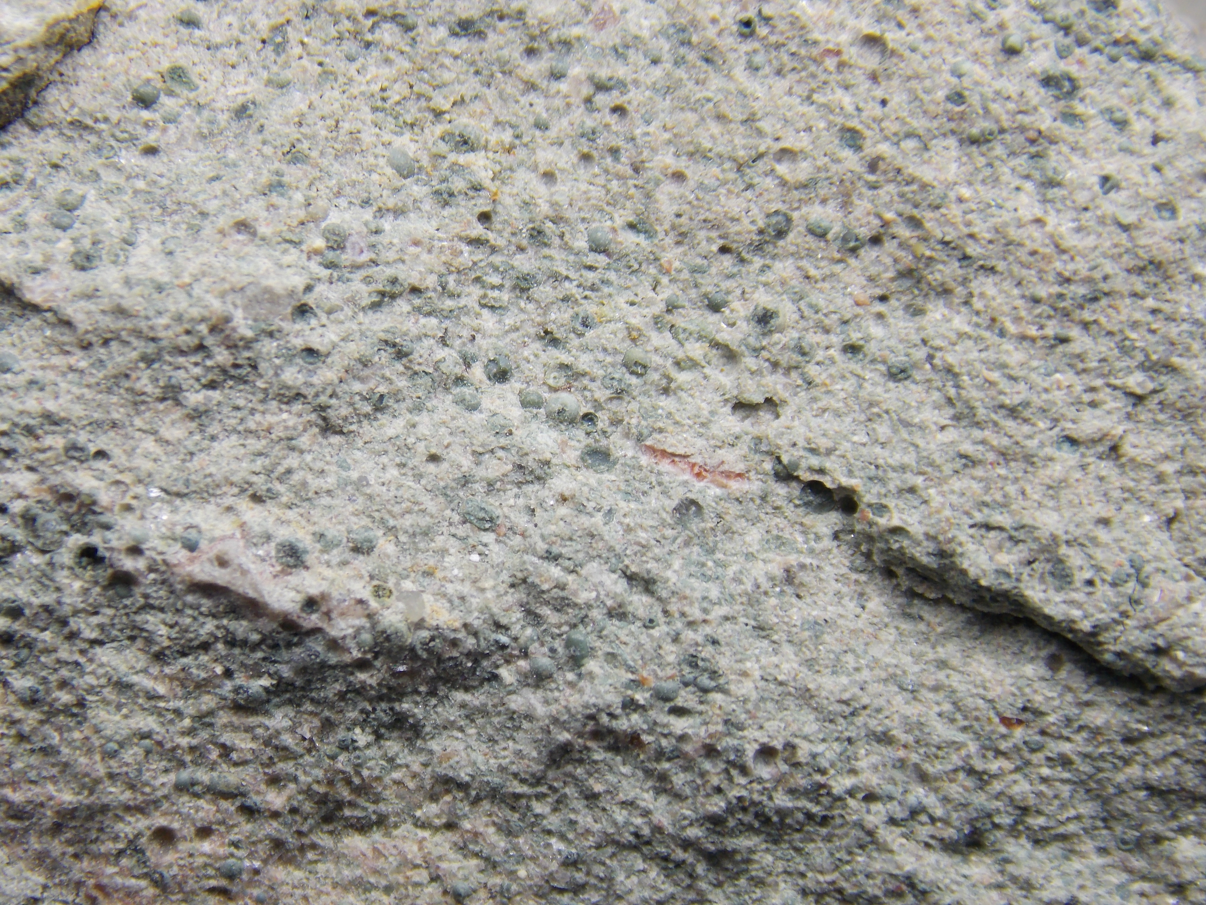
Field of view: 30 x 22mm
The spherules can clearly be seen interspersed with angular
grains of carbonate and occasional quartz. It is not
possible to determine the nature of the spherules in this
view but they lack the vitreous lustre expected of silica.
|
|
Thin section
|
Thin section in plane polarised light (full section)
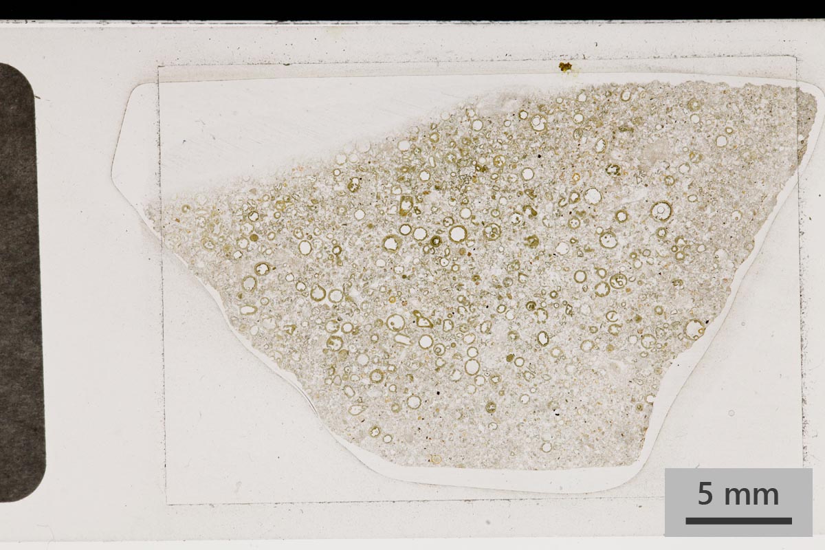
PPL view. Small green-brown spherules are visible in this
view. These are interpreted as micro-tektites from a meteor
impact which have been reworked into a small cross-bedded
channel fill. Some quartz grains are present.
|
Thin section in cross polarised light (full section)
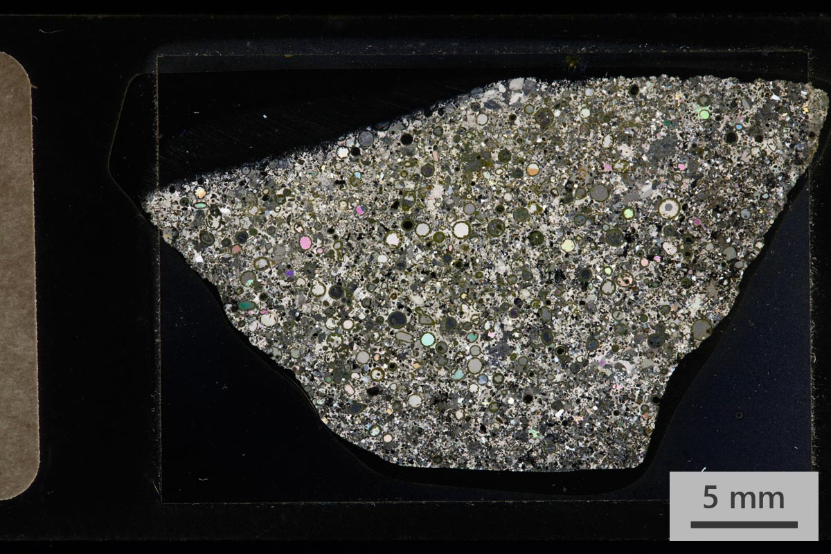
XPL view. The centres of the micro-tektites have been filled
with either a single calcite crystal or 2-3 large crystals.
The grains have been cemented with sparry calcite which may
have partially replaced an original finer marl (carbonate
mud). Some large calcite crystals have grown around other
grains to create a poikilitic texture.
|
Thin section, detailed
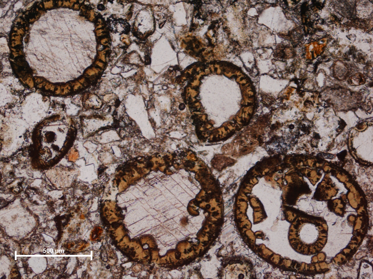
In this plain polarised light view details of the
micro-tektites can be seen more clearly. The walls of the
spherules are glauconitic in nature but would have
originally been silica glass. The proposed process for the
formation of the glauconite is that the original silica was
hydrated to form a silica gel (palagonitization), this was
then altered to smectite and finally to glauconite
(glaconitization). Studies have not been able to prove this
is true glauconite so instead the term glauconitic is used
to imply it has a chemistry and crystal structure similar to
glauconite. Growth of the replacement glauconitic material
appears to have propagated inwards as demonstrated by the
botryoidal shapes on the inner surfaces. The spherule to the
top left is the most perfectly formed. Slightly above right
of centre is a deformed spherule which may have been damaged
as it was redeposited from its original location. The
spherule below left of centre clearly demonstrates the
botryoidal nature of the glauconitic material. A spherule
containing an intact smaller spherule and fragments of a
broken spherule can be seen bottom right.
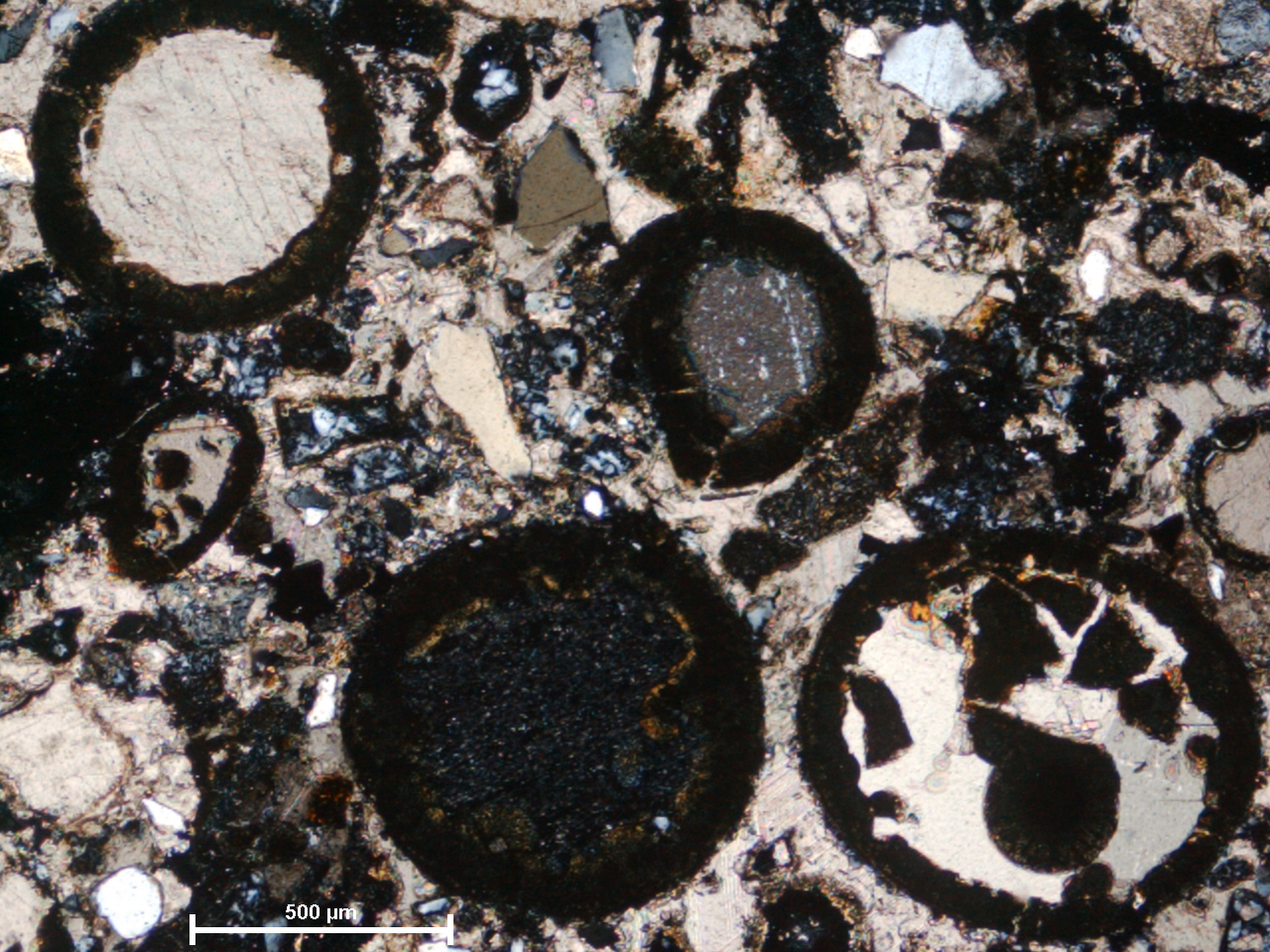
The same view under crossed polars shows single calcite
crystals filling the three spherules furthest left. The
multiple spherule to the bottom right contains 2 calcite
crystals, though this is hard to determine in this view.
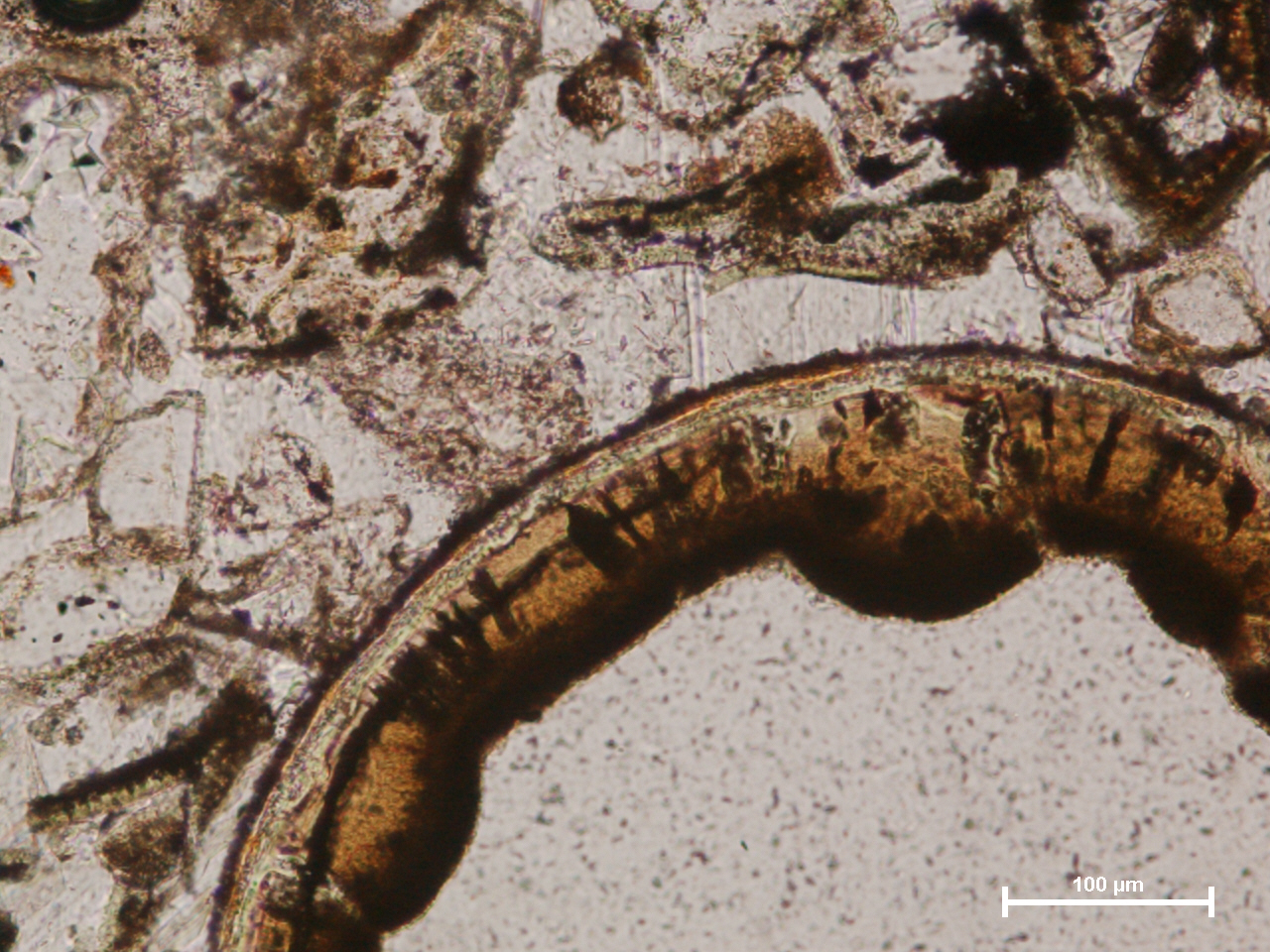
In this plain polarised light view at higher magnification
the shell of the spherule can be seen more clearly. This
more perfect spherule is unfilled; the textured material
within it is resin. The thin outer surface of the spherules
has been identified as sericitic though this cannot be
proven by optical microscopy. The thicker glauconitic layer
shades through to dark brown towards the centre of the
spherule due to increasing iron content.
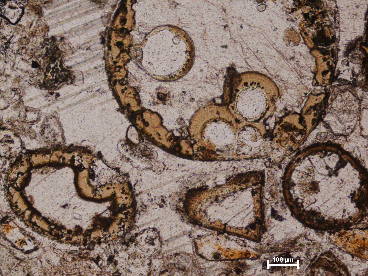
Further examples are demonstrated here in plane polarised
light. In the large spherule to the top of the view several
internal spherules can be seen. It is interesting to note
that the growth pattern is reversed on internal spherules,
i.e. the sericitic layer is closest to the inside of the
spherule, followed by a thick glauconitic shell with
increasing iron content towards the outermost surface. The
spherule to the bottom left is heavily embayed, possibly due
to damage to the spherule before it was glauconitized. An
oddly shaped rhombic “spherule” can be seen lower right of
centre. Though most of the spherules are relatively intact
some heavily distorted examples such as this do occur. Note
that a large single calcite crystal envelopes most of the
grains on the left hand side of the image giving a
poikilitic texture.
|